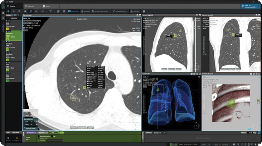
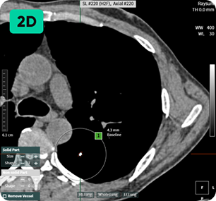
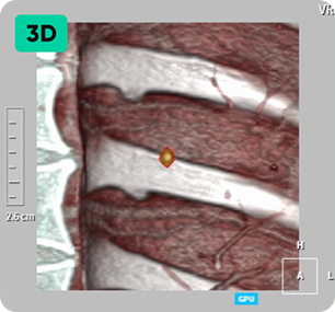
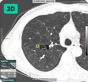
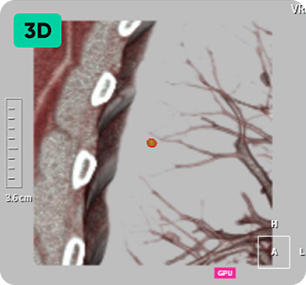
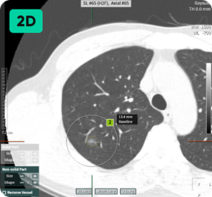
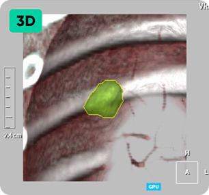
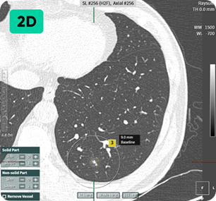
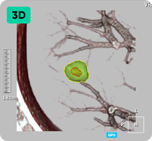
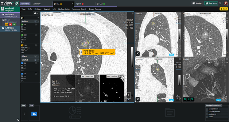
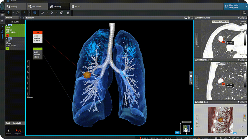
GC&Phenikaa

0961 580 001

Giải pháp AI
của chúng tôi

Đi đầu trong đổi mới, giải pháp chăm sóc sức khỏe ứng dụng AI của chúng tôi tận dụng công nghệ tiên tiến để chuyển đổi ngành y tế. Bằng cách tích hợp học máy, phân tích dự đoán và các thuật toán hiện đại, giải pháp này được thiết kế nhằm nâng cao chất lượng chăm sóc bệnh nhân, tối ưu hóa hoạt động bệnh viện và hỗ trợ các chuyên gia y tế đưa ra quyết định một cách chính xác.

LOGO CORELINE-GC&Phenikaa
LOGO LUNIT-GC&Phenikaa
LOGO WAYCEN-GC&Phenikaa
1

Automated Nodule Detection
and Lung-RADS Calculation

Detecting and Analyzing Easy to miss Lung Nodules on Low-Dose Chest CT with Imaging Artificial Intelligence.

Efficiently diagnose with awareness of patient conditions.

Finding microscopic nodules provides a variety of information, including basic, number of nodules, size and status, and RADS category. Findings that are likely to develop into lung cancer can also be checked in advance, reducing working time and allowing efficient reading depending on the case. Microscopic Nodule Detection: Providing Comprehensive Information on Number of Nodules, Size, Status, and RADS Category Early Detection of Potential Lung Cancer Development Enables Time-efficient Scans Reading.

Detecting Nodules of Various Sizes, from Small to Large

anh-c-ai-1 anh-c-ai-h1
anh-c-ai-2 anh-c-ai-h2
anh-c-ai-3 anh-c-ai-h3
anh-c-ai-4 anh-c-ai-h4
anh-c-ai-5 anh-c-ai-h5
2

Follow up mode

Automated Matching of Follow-up with Previous Lung CT Scans Instantly Assess Nodule Changes.

Monitoring Nodule Growth is Vital for Accurate Readings.

It autonomously assesses growth and changes by matching lung nodules, going beyond mere image comparisons.
aview LCS efficiently classifies lung nodules into the relevant categories according to the Lung CT Screening Reporting and Data System (Lung-RADS ver 1.1) guidelines as recommended by the American College of Radiology.

Follow-up CT Scan

Previous CT Scan

3

Clinical Viewer

CT Findings are visualized in 3D.
User-Friendly Viewer for Medical Professionals and Patients.

Advanced 3D Rendering of Patient’s Lung.

2D Images May Not Provide Adequate Information of Abnormal Findings Visualized 3D Model of Patient’s Lung is much more informative and intuitive. Abnormal findings can be easily explained by demonstration of the 3D Model.