GC&Phenikaa

Our AI Solutions

At the forefront of innovation, our AI-powered healthcare solution leverages cutting-edge technology to transform the healthcare industry. By integrating machine learning, predictive analytics, and advanced algorithms, it is designed to improve patient care, optimize hospital operations, and empower healthcare professionals to make informed decisions.

LOGO CORELINE-GC&Phenikaa
LOGO LUNIT-GC&Phenikaa
LOGO WAYCEN-GC&Phenikaa
Công nghệ AI
1

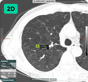
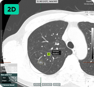
Automated Nodule Detection
and Lung-RADS Calculation

Detecting and Analyzing Easy to miss Lung Nodules on Low-Dose Chest CT with Imaging Artificial Intelligence.

Efficiently diagnose with awareness of patient conditions.

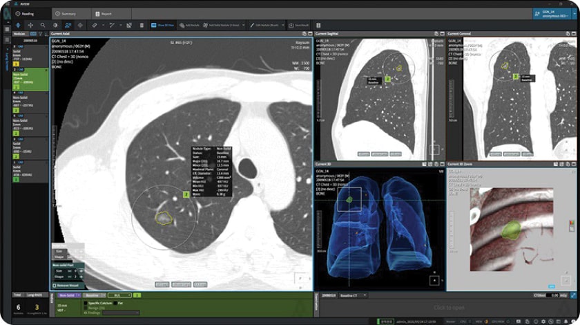
Finding microscopic nodules provides a variety of information, including basic, number of nodules, size and status, and RADS category. Findings that are likely to develop into lung cancer can also be checked in advance, reducing working time and allowing efficient reading depending on the case. Microscopic Nodule Detection: Providing Comprehensive Information on Number of Nodules, Size, Status, and RADS Category Early Detection of Potential Lung Cancer Development Enables Time-efficient Scans Reading.


Rectangle 18

Detecting Nodules of Various Sizes, from Small to Large

Công nghệ AI Công nghệ AI
2

Follow up mode

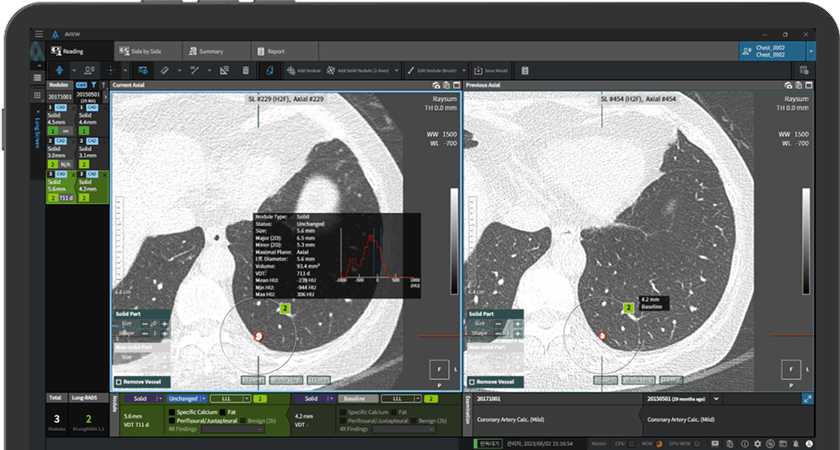
Automated Matching of Follow-up with Previous Lung CT Scans Instantly Assess Nodule Changes.

Monitoring Nodule Growth is Vital for Accurate Readings.

It autonomously assesses growth and changes by matching lung nodules, going beyond mere image comparisons.
aview LCS efficiently classifies lung nodules into the relevant categories according to the Lung CT Screening Reporting and Data System (Lung-RADS ver 1.1) guidelines as recommended by the American College of Radiology.

Follow-up CT Scan

Previous CT Scan

sol02new 1
3

Clinical Viewer

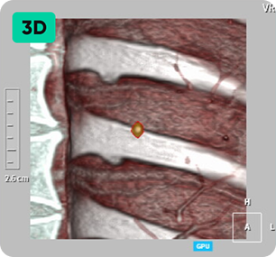
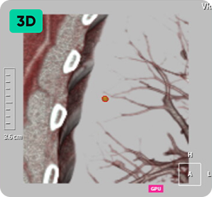
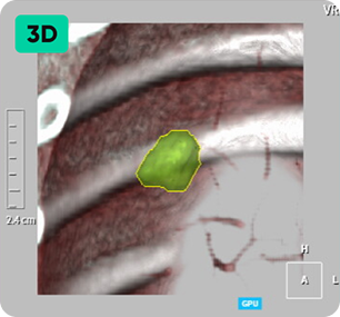
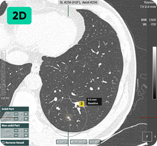
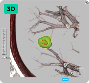
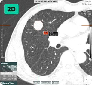
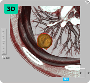
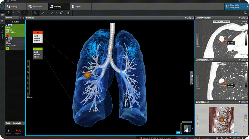
CT Findings are visualized in 3D.
User-Friendly Viewer for Medical Professionals and Patients.

Advanced 3D Rendering of Patient’s Lung.

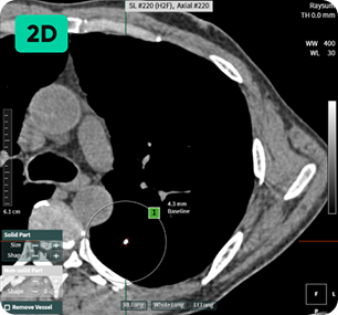
2D Images May Not Provide Adequate Information of Abnormal Findings Visualized 3D Model of Patient’s Lung is much more informative and intuitive. Abnormal findings can be easily explained by demonstration of the 3D Model.

Công nghệ AI